


Contributing € 10
ACKNOWLEDGEMENT
Personal acknowledgement in the group web site.
> 13 Backers
We use own and third party cookies to improve your user experience and our services, analyzing users' browsing in our website. If you continue browsing, we will consider that you consent to its use. You can get further information in our Cookies Policy
Herramienta diagnóstica en leucemias y linfomas
 Barcelona


AUTOMATIC CLASSIFICATION OF DIGITAL IMAGES OF PERIPHERAL BLOOD: APPLICATION TO THE INITIAL DIAGNOSIS OF LEUKEMIAS AND LYMPHOMAS
 Task
Task
|
Minimum | Optimum |
|---|---|---|
|
Digital microscopy camera
N4257100
DP73-1-51 Digital Microscope Camera.
It is a professional colour digital camera for light microscopy. It allows captures with an effective resolution up to 4800x3600 píxels. A new capture mode that allows to obtain real colour images without interpolation. It includes the technology Fine Detail Process, which increases the brightness in the smallest details.
|
€ 8.950 | |
|
PROGRAM FOR IMAGES MANAGEMENT
N5157500
CS-DI-V1.8 cellSens Dimension V1.8.
Image capture and analysis program for applicacions in microscopy in life sciences. It includes basic tools as information display and image properties, virtual navigator, view of active images gallery and combination of RGB images in a single image. It also allows the image processing through filters and other tools, as well as the possibility of making notes on the image.
It has extended functions like multidimensional image processing and image geometry. Image filters to improve contrast, smoothing or roughness increase and homogeneization of image background.
|
€ 3.000 | |
|
MICROSCOPE FOR RESEARCH APPLICATIONS
N2709900
BX53F versatile microscope frame for research applications.
The Koehler lighting system has a halogen lighting power of 100 W, keeping the temperature constant for an optimal colour reproduction in the image capture. It integrates a grading filter that allows full homogeneity in the lighting field for any magnification.
Moreover, it incorporates a set of filters LBD (Light Balance Daylight) allowing an ideal white light for the reproduction of colour in clear field in the whole observation field and neutral filters for the intensity control.
|
€ 5.500 | |
| Total | € 11.950 | € 17.450 |
Severe hematological diseases, especially acute leukemias and lymphomas, are common in all ages of life and its frequency increases with age. Detection and early treatment are essential to the survival of the patients, as they constitute one of the leading causes of death in the population.
Our project aims to develop a new tool to support the detection of these serious blood diseases such as leukemia and lymphoma. In the last two years a collaboration was initiated between the Technical University of Catalonia (UPC) and the Hospital Clínic of Barcelona for the joint development of mathematical tools to aid initial diagnosis of leukemia from its application in microscope digital images of blood cells.
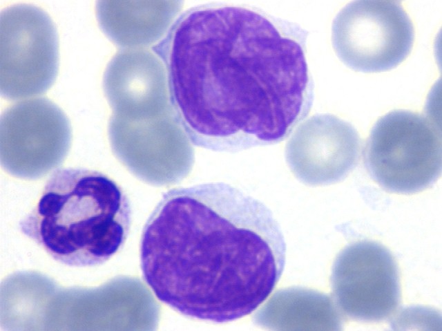
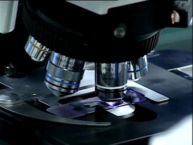
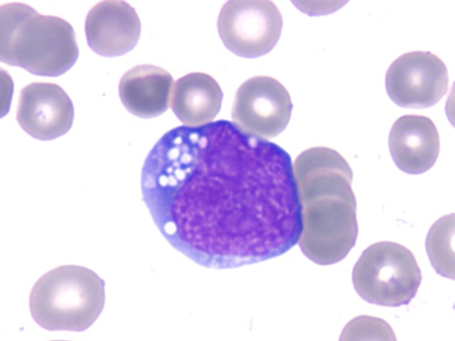
Blood is a fluid very easy to obtain. The study of the characteristics of the blood cells under the microscope provides very useful information, and is the first analytical step in diagnosing leukemias. In our project we will work with high-resolution digital images obtained from a drop of blood stained with dyes, and acquired by a camera coupled to a microscope.
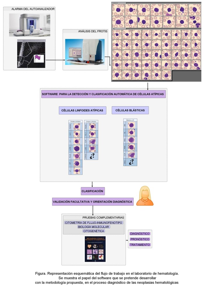
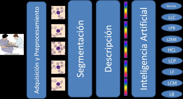
We will try to classify automatically malignant blood cells, combining the knowledge of the skilled clinician with new artificial intelligence methods.The ultimate goal of the project is to design software tools to implement the classification method and allow its use in clinical practice as a diagnosis support tool. In order to do this, the following steps are proposed:
1) Creation of a broad base of digital images obtained using a microscope to extract the morphological characteristics of the cell types under study;
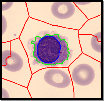
2) development of a strategy for the preprocessing of the images;
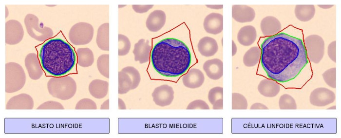
3) use of a robust method for segmenting images to separate malignant cells from the remaining components of the image obtaining different regions (nucleus, cytoplasm and outer region);
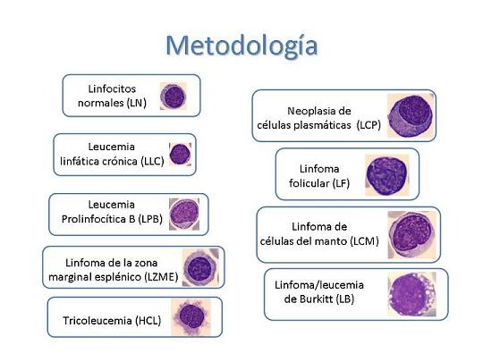
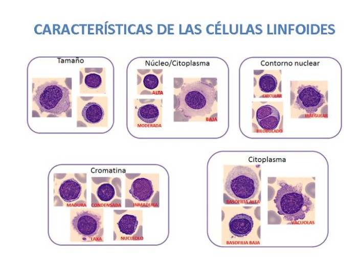
4) definition of descriptors well suited for the identification of different cell types;
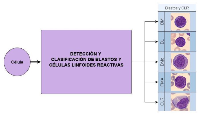
5) classification of digital images of leukemic cells using artificial intelligence techniques;
6) development of a software in order to implementate the methodology.
The project will be developed in two years.
The development of practical algorithms and their implementation through software systems can be a valuable support for diagnosis, allowing to obtain objective and reproducible values in the interpretation of images of leukemic cells. The development and implementation of the proposed methodology can be a breakthrough for automated classification of leukemia cells that are observed in the blood for different diseases. It may also contribute to a better use of other tests, complex and with high cost, which are made only in highly specialized laboratories.
The proposed project is important for the quick diagnosis of the patient and subsequent treatment, which will positively influence the prognosis. We combine a mathematically based research with the expertise of medical professionals who know real and practical needs. We are motivated by a common goal: to obtain useful and cost-effective products that help to improve the diagnosis. We share the complicity of both the UPC and the Hospital Clínic, two major institutions in their areas of care, teaching and research. We are particularly motivated by going out of our laboratory doors and involving society in our work and in our progress.
We address to all those professionals, patients and people in general to consider the possibility of becoming involved in our exciting project.
The challenge of our project is to design a new method that helps the quick diagnosis of leukemias and lymphomas. Our hope is that this campaign of microfinance, beyond concrete support for this project, will serve to boost and support for future projects in this research line of biomedical engineering, which we intend to benefit the people.
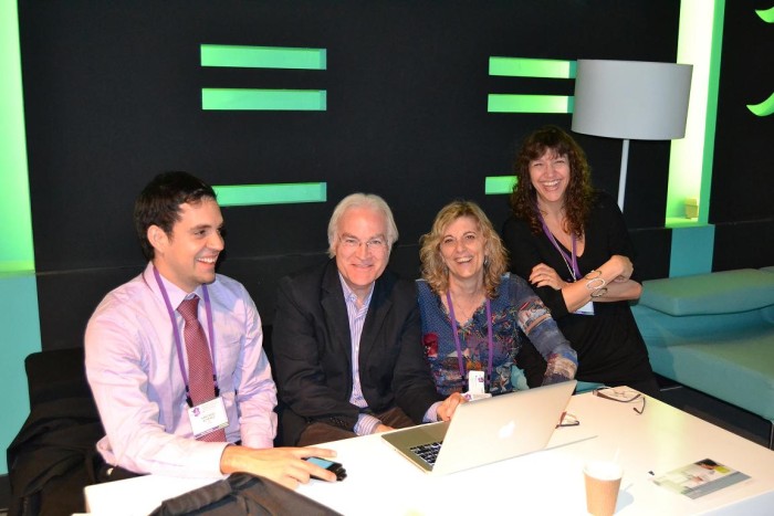
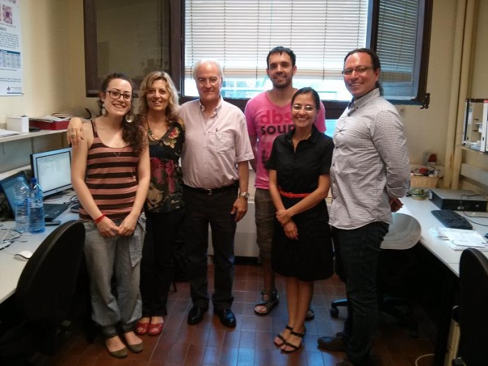
This project is carried out within a multidisciplinary team of researchers from the CoDAlab group, led by José Rodellar, Professor, Department of Applied Mathematics III of the UPC and Dr. Anna Merino, Cytology Consultant of Hospital Clínic and researcher in Oncology and Hematology at IDIBAPS.
The collaboration began in 2010 with a project on applications of digital image processing of peripheral blood, framed on the completion of the doctoral thesis of Santiago Alferez, within the program of Biomedical Engineering of the UPC, and directed by Dr. Rodellar and Dra. Merino. Since then, we have two more engineering researchers from UPC, Dr. Luis Mujica and Dr. Magda Ruiz, working with us in this project. In 2012 we have in the group a post-residency fellowship from the Foundation JL Castaño, Laura Bigorra, Clinical Chemistry analysis specialist, which, at present, is also developing her PhD in Biomedical Engineering Program at the UPC.
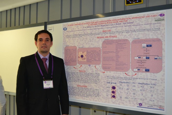
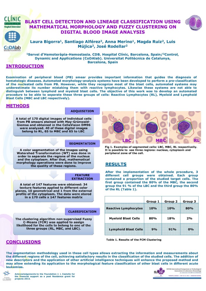
The team presented results at international conferences of ISLH (International Society of Laboratory Hematology): "Morphological features microscopic blood analysis by segmentation using digital image processing in lymphoid neoplasms", "Atypical lymphoid cells detection and classification using mathematical morphology on blood digital images", "Blast cell lineage detection and classification using mathematical morphology and fuzzy clustering on digital blood image analysis" and " Atypical lymphoid cells detection and classification on blood digital image analysis ". We are going to publish our preliminary results in the International Journal of Laboratory Hematology. One of the comments of the reviewers about our article was: "This is an interesting article that will contribute to further develop and improve the automated classification of peripheral blood cells"
With the method and preliminary results, the group has submitted (may 9, 2013) the Spanish request for patent with No. P201330671, with prior agreement Clínic-UPC, entitled: "Computer implemented method for the recognition and classification of abnormal blood cells and software to perform the method".


Ensuring healthy lives and promoting the well-being for all at all ages is essential to sustainable development.